1. Why Does a Dental Technician Need Radiographic Awareness?
Radiographs have become increasingly visible within the dental laboratory. A technician may receive a periapical radiograph alongside a crown or bridge case, an OPG as part of a complex removable case, or a CBCT dataset as part of an implant or guided-surgery workflow. In digital dentistry, DICOM data may also be combined with intraoral scans, facial scans and digital photographs.
This does not mean that every technician becomes a radiographic diagnostician. The purpose of radiographic awareness in the laboratory is different. A technician needs to understand what information an image can provide, recognise obvious limitations, communicate effectively with the prescribing clinician and understand when a question belongs back with the clinical team.
The GDC Scope of Practice is particularly important here. The current guidance makes clear that dental professionals must work within the boundaries of their professional title and their own personal scope of practice. A task may be technically possible but still be inappropriate if the individual does not have the necessary training, competence, confidence or indemnity.
There is also an important distinction between dental technicians and clinical dental technicians. Clinical dental technicians can, when appropriately trained and competent, prescribe, undertake and interpret radiographs within their scope. That should not be confused with the role of a dental technician who is using radiographic information supplied by a prescribing clinician as part of laboratory work.
For the laboratory technician, therefore, radiographic awareness is primarily about informed technical communication: understanding the information, recognising limitations and knowing when the clinical team needs to make the decision.
2. Reading the Image: Radiopaque, Radiolucent and Anatomical Information
A useful starting point for radiographic awareness is understanding the basic language used to describe appearances. Structures that attenuate more X-rays generally appear more radiopaque, while structures that allow more radiation through appear more radiolucent.
Dental materials can produce particularly strong radiopaque appearances. Metals, zirconia and other high-density materials may appear very bright on a radiograph. Enamel and dentine are also relatively radiopaque, while pulp spaces, periodontal ligament spaces and many soft-tissue structures appear comparatively radiolucent.
Technicians working with restorative and implant cases may encounter familiar anatomical structures such as the mandibular canal, mental foramen, maxillary sinus and nasal cavity. Recognising these structures can be useful when communicating about a case or understanding the information provided by a clinician.
However, recognising an anatomical structure is not the same as diagnosing a condition. For example, a technician may recognise that an area appears unusual or that an anatomical boundary is difficult to identify, but deciding whether that appearance represents pathology, anatomical variation or an imaging artefact is a clinical interpretation.
This distinction is particularly important when radiographs are received as part of a laboratory prescription. The technician should not assume that the laboratory is being asked to independently determine whether a patient is clinically suitable for treatment. Where clinical suitability or diagnosis is relevant, that responsibility remains with the appropriately trained clinical professional.
Radiographic awareness therefore supports better communication. A technician who understands the image can ask a more useful question: “The mandibular canal is difficult to identify in this region of the CBCT; could you confirm the intended planning information before we proceed?” That is very different from independently deciding where an implant should be placed.
3. CBCT: Three Dimensions, More Information — and More Complexity
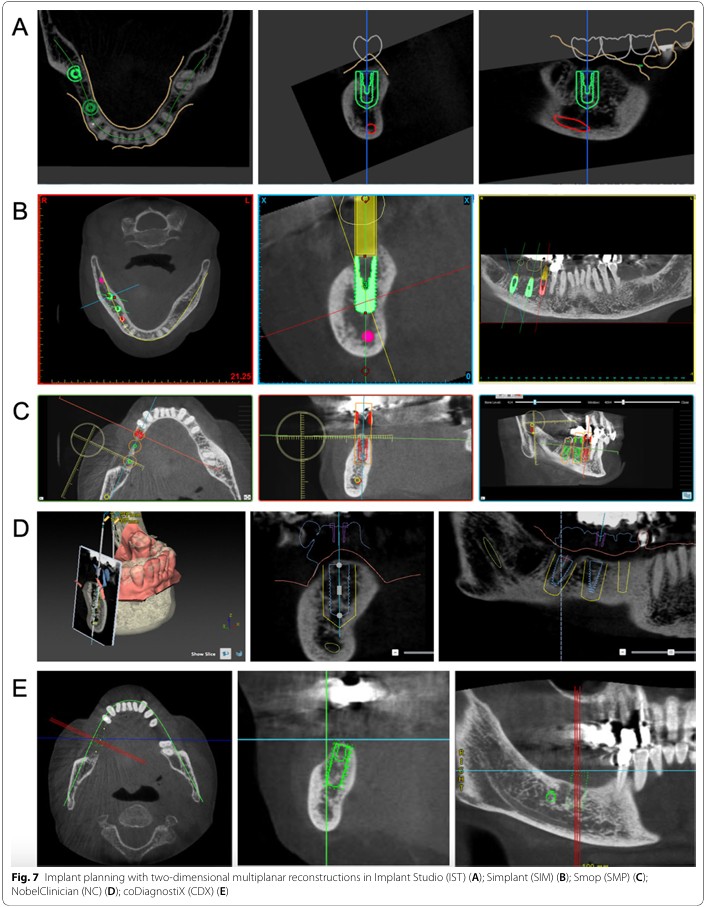
Cone Beam Computed Tomography provides a three-dimensional dataset that can be viewed in multiple planes. Depending on the software, the technician may work with axial, sagittal and coronal views alongside reconstructed panoramic and cross-sectional views.
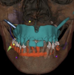
For digital implant and surgical workflows, CBCT data may be combined with an intraoral scan. This allows the laboratory to relate the proposed prosthetic design to the three-dimensional information contained within the radiographic dataset.
However, CBCT should not be treated as a perfect three-dimensional representation of the patient. Image quality depends on factors including the acquisition parameters, voxel size, patient movement, positioning and the presence of high-density materials.
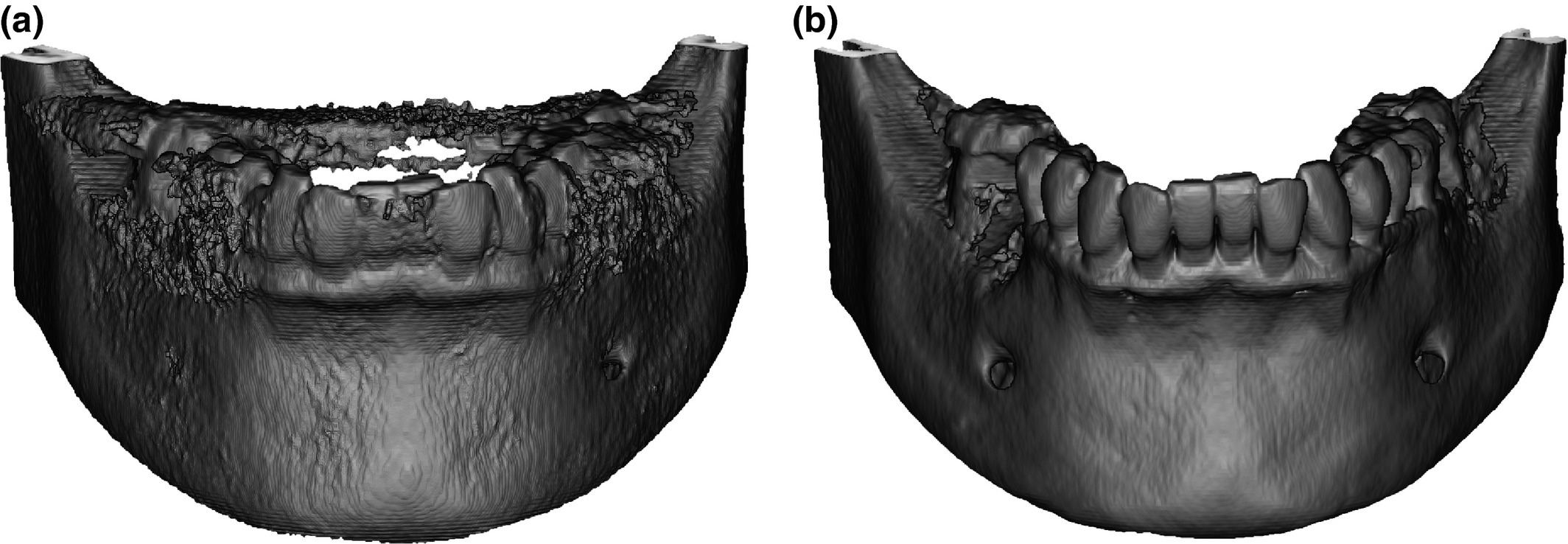
The laboratory should also be aware of the difference between the original radiographic dataset and the surfaces generated from it. Segmentation software creates a representation from the underlying data; it does not remove the limitations of that data. An apparently clean digital surface may therefore give a false impression of certainty if the original CBCT contains significant artefact.
In a guided-surgery workflow, this distinction matters. The technician may be responsible for the technical design and manufacture of a guide based on information provided by the clinical team, but should not silently compensate for uncertainty in the radiographic data by making independent clinical assumptions.
If the dataset is incomplete, corrupted, poorly matched or contains areas that cannot be interpreted reliably for the intended workflow, the appropriate response may simply be to stop and seek clarification.
4. Artefacts: When the Image Is Not Telling the Whole Story
One of the most important aspects of radiographic awareness is understanding that not every feature visible on an image represents a real anatomical structure.
Metallic restorations are a common source of CBCT artefact. High-density materials can produce streaking, areas of increased or reduced apparent density and other distortions around the restoration. These effects can obscure anatomical boundaries and interfere with segmentation or registration.
Patient movement can produce another important limitation. Movement during acquisition may result in blurred or duplicated structures, making the relationship between teeth, bone and other anatomy less reliable.
Other limitations can arise from incomplete fields of view, poor positioning, inadequate resolution or grey settings within the dataset itself. A technician working with digital planning software should learn to recognise when the image is behaving unexpectedly rather than assuming that every visible boundary is accurate.
Artefact awareness is particularly relevant when registering CBCT data with an intraoral surface scan. A technician may be tempted to force two datasets to align because the software requires a successful registration. However, a mathematically successful registration is not necessarily a clinically meaningful one.
Where artefact prevents reliable interpretation of the information needed for the laboratory workflow, the correct response is not to guess. The technician should communicate the limitation to the prescribing clinician and seek appropriate clarification or replacement information.
5. Radiographic Information in Implant and Restorative Workflows
Implant dentistry provides some of the clearest examples of how radiographic information and laboratory design interact. A clinician may provide a CBCT dataset alongside an intraoral scan, photographic records and a restorative prescription. The laboratory may then use these datasets to develop a prosthetically driven design or manufacture a surgical guide.
The important professional boundary is that the technician is working from information provided as part of the clinical planning process. The technician may identify whether the datasets appear to align, whether the proposed design can be manufactured and whether the digital workflow is technically achievable.
The technician should not, however, independently decide that an implant is clinically appropriate, diagnose bone pathology, determine whether augmentation is required or prescribe a surgical trajectory simply because the software makes these actions possible.
Where a design reveals an apparent conflict — for example, a proposed implant position appears incompatible with the restorative design, available space or information provided by the clinician — this is an opportunity for communication rather than unilateral clinical decision-making.
This is where the laboratory can add considerable value. A technician who understands the relationship between the prosthetic design and the radiographic information can identify technical or restorative conflicts early and communicate them clearly to the clinical team.
The strongest digital workflows are therefore collaborative. The clinician provides the clinical diagnosis, treatment plan and relevant radiographic interpretation. The technician contributes specialist knowledge of prosthetic design, manufacturing, materials and digital workflow. Each professional works within their own scope while communicating effectively with the rest of the team.
6. Knowing the Boundary: Observation, Interpretation and Diagnosis
The most important lesson in radiography awareness for dental technicians is knowing where technical observation ends and clinical interpretation begins.
Consider three statements: “The mandibular canal is difficult to visualise in this section.” “There may be insufficient bone for the proposed implant.” “The patient has significant bone loss.” The first is a technical observation. The second begins to express a clinical planning judgement. The third is a clinical interpretation or diagnosis. They are not interchangeable.
The GDC’s current Scope of Practice places considerable emphasis on professional judgement, competence and boundaries. Dental professionals must only carry out tasks for which they are appropriately trained, competent and indemnified or insured. If a task falls outside their scope or competence, they should not simply proceed because the technology makes it possible.
For technicians, this is especially relevant as digital systems become increasingly sophisticated. CAD software can display nerve canals, identify anatomical structures, generate implant proposals and provide measurements. The availability of a tool does not automatically confer professional authority to make the decisions that the tool enables.
A technician can therefore add significant value without becoming the clinician. They can identify technical limitations, question incomplete information, recognise obvious artefacts, check whether datasets are compatible and communicate restorative or manufacturing implications.
The professional skill is knowing when to say: “I can identify this technical issue, but the clinical decision needs to come from the prescribing clinician.” That is not a limitation of expertise. It is evidence of appropriate professional judgement.